
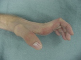
If the median nerve is injured as well, the thumb may be unable to be drawn out of the plane of the palm--making grip and pinch nearly impossible. A claw deformity look like this:
Intrinsic paralysis results in MP joint hyperextension. A claw deformity may be worse with a low ulnar nerve injury since the profundus tendons still function, and will exacerbate the claw. Because reciprocal deformity results in PIP joint flexion, the lateral bands drop volar and extension of the IP joints is prohibited, unless the MP joints are held in flexion---this then allows the extrinsic extensor to extend the PIP joint via the central slip. This finding is called a Positive Bouvier test. If the test is Negative---with the MP joints held in flexion, the IP joints still can't extend-- then tendon transfers ideally will be performed to restore intrinsic function to the IP joints via a connection to the lateral bands, as opposed to the proximal phalanx alone (which is done with a dynamic intrinsic transfer to eradicate clawing when the Bovier test is positive.)
The fundamental goals of tendon transfer surgery for Ulnar nerve palsy and Combined palsy include improving pinch and grip strength, and correcting claw deformity. This facilitates the restoration of synchronous finger flexion--initiated at the MP joints.
Videos below show:
- Asynchronous finger flexion and claw deformity that result from intrinsic paralysis. The fingers curl on themselves because flesion is initiated at the tips from the FDP.
- A positive Bouvier test, indicating adequate finger IP extension when the MPJ hyperextension is corrected.
- 2 videos of a patient who had a combined median and ulnar palsy reconstructed via transfers: Brachioradialis to FPL, EIP opponensplasty, APL to 1st DI, and ECRL to the finger intrinsics, volar to the axis of rotation.
- Patient AD who had a combned median and ulnar palsy and will underwent transfers to address deficient thumb opposition. claw deformity, and a negative Bouvier test.
Patient AD had sustained lacerations of radial, ulnar, and median nerve injuries in the upper arm 20 years earlier, at which time nerve repair was permed. She recovered some median and radial function and had good FDP and FDS function but no opposition. She also recovered grade 5 finger extension and wrist extension.
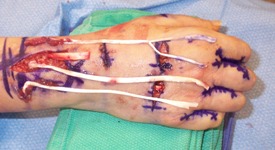
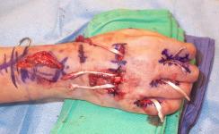
Ring finger FDS opponens transfer ws performed. The FDS is held in the clamp after passage.
.jpg)
Bilateral Palmaris longus and small finger FDS were used as graft to correct claw deformity and improve PIPJ extension as described by Brand. The ECRL is the motor. The grafts are passed volar to the deep transverse metacarpal ligament and are sewn to the radial lateral bands.

Throughout my career, I have had the priviledge of performing reconstructive surgery for ulnar nerve palsy and combined palsy of the medial and ulnar nerves in over 30 patients. This experience, and my commitment to scrutinize postoperative outcomes, has afforded me a unique opportunity to assess functional return, levels of patient satisfaction, and to constantly improve surgical techniques and selection thereof.
Though the hand will never work normally, surgery is very effective at correcting claw deformity, restoring synchronous finger flexion,improving the position of the thumb, and improving prehensile pinch strength between thumb and index finger. When there is an element of median nerve palsy, the tendon transfer to the thumb can be altered a bit to improve the ability to position the thumb in space, in preparation for grip.
Surgery takes approximately 2-3 hours and can be performed as an outpatient. Tendon grafts are usually required, but donor tendons from either arm or feet are available--without residual loss of function. After surgery a cast is worn for 4 weeks, followed by a removable splint for 4 weeks. Strengthening begins at 8 weeks, and full recovery may take as long as 6-8 months.